42 easy microscope diagram with labels
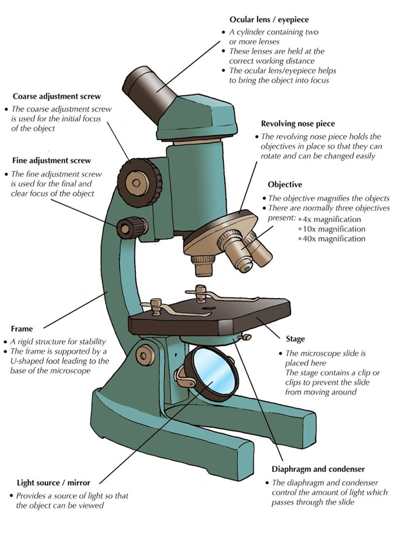
Microscope, Microscope Parts, Labeled Diagram, and Functions Revolving Nosepiece or Turret: Turret is the part of the microscope that holds two or multiple objective lenses and helps to rotate objective lenses and also helps to easily change power. Objective Lenses: Three are 3 or 4 objective lenses on a microscope. The objective lenses almost always consist of 4x, 10x, 40x and 100x powers. The most common eyepiece lens is 10x and when it coupled with ... › publication › 320945390(PDF) Introduction to Microscopy - ResearchGate Nov 08, 2017 · 1. Microscopy with light and electrons 2. Electron/specimen interactions: processes and detectors 3. The electron microscope family 4. Specimen preparation for electron microscopy 5.
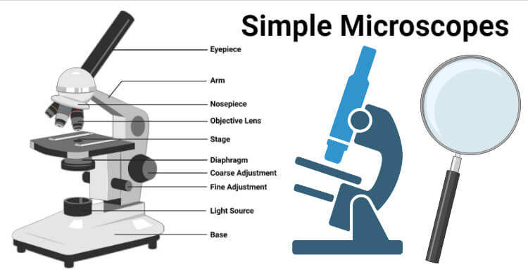
Compound Microscope Parts - Labeled Diagram and their Functions The term "compound" refers to the microscope having more than one lens. Basically, compound microscopes generate magnified images through an aligned pair of the objective lens and the ocular lens. In contrast, "simple microscopes" have only one convex lens and function more like glass magnifiers. [In this figure] Two "antique ...
Easy microscope diagram with labels
Parts of a Simple Microscope - Labeled (with diagrams) image 5: A modern simple microscope with the different parts labeled. image source: laboratoryinfo.com The optical parts of a simple microscope are centered on the specimen - lighting, and magnification. Label the Microscope Diagram | Download Scientific Diagram - ResearchGate Gram staining was performed using compound microscope according to the procedure described by Petersen et al., 2016 [8]. The gram positive and gram negative bacteria were identified based on ... Microscope With Labels clip art | Microscope parts, Scientific method ... Description Use this blank handout as a way for students to record microscope drawings. Aside from the drawig itself, studnts are prompted to title the drawing, include the magnification of the microscope, and give a quick description of what they are viewing. Keywords: Microscope Drawings Lab Biology. S. Learning with Shedd Aquarium.
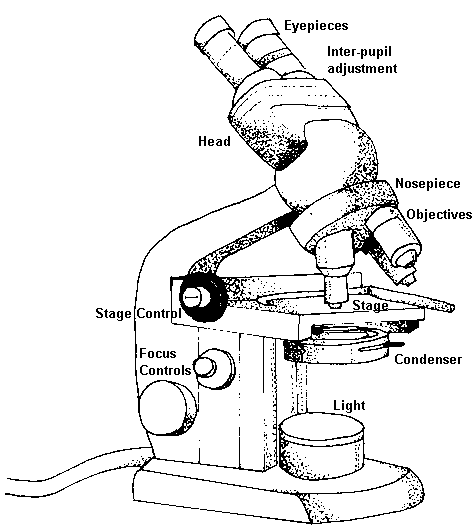
Easy microscope diagram with labels. rsscience.com › stereo-microscopeParts of Stereo Microscope (Dissecting microscope) – labeled ... Labeled part diagram of a stereo microscope Major structural parts of a stereo microscope. There are three major structural parts of a stereo microscope. The viewing Head includes the upper part of the microscope, which houses the most critical optical components, including the eyepiece, objective lens, and light source of the microscope. Compound Microscope Parts, Functions, and Labeled Diagram Compound Microscope Definitions for Labels. Eyepiece (ocular lens) with or without Pointer: The part that is looked through at the top of the compound microscope. Eyepieces typically have a magnification between 5x & 30x. Monocular or Binocular Head: Structural support that holds & connects the eyepieces to the objective lenses. Simple Microscope - Diagram (Parts labelled), Principle, Formula and Uses Simple microscope is a magnification apparatus that uses a combination of double convex lens to form an enlarged, erect image of a specimen. The working principle of a simple microscope is that when a lens is held close to the eye, a virtual, magnified and erect image of a specimen is formed at the least possible distance from which a human eye ... › newgrouppage9Epi-Illumination for DIY Cerna® Systems - Thorlabs Apr 01, 2022 · The Cerna microscopy platform's large working volume and system of dovetails make it straightforward to connect and position the components of the microscope. This flexibility enables simple and stable set up of a preconfigured microscope, and provides easy paths for later upgrades and modification.
The Parts of a Microscope (Labeled) Printable Printable (6th - 12th ... The Parts of a Microscope (Labeled) Printable. Download. Add to Favorites. Share. This diagram labels and explains the function of each part of a microscope. Use this printable as a handout or transparency to help prepare students for working with laboratory equipment. Grade: › articles › nprotGeneration of cerebral organoids from human pluripotent stem ... Sep 04, 2014 · Size measurements can be performed using an inverted microscope equipped with a camera and measurement software. Typically, ROCK inhibitor and low-bFGF medium are included only for the first 4 d. 4 Label the microscope — Science Learning Hub All microscopes share features in common. In this interactive, you can label the different parts ... Microscope Labeling Game - PurposeGames.com About this Quiz. This is an online quiz called Microscope Labeling Game. There is a printable worksheet available for download here so you can take the quiz with pen and paper. This quiz has tags. Click on the tags below to find other quizzes on the same subject. Science.
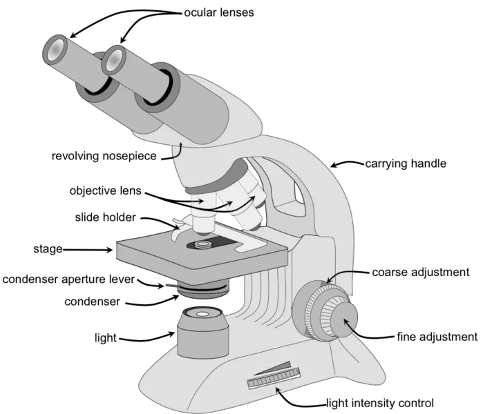
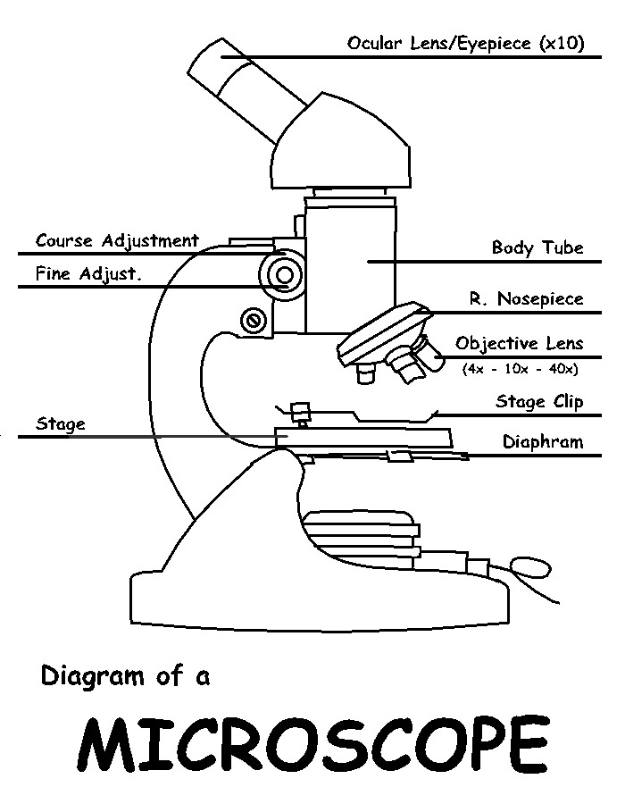
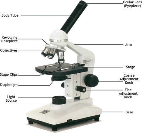
Label Microscope Diagram - EnchantedLearning.com Using the terms listed below, label the microscope diagram. arm - this attaches the eyepiece and body tube to the base. base - this supports the microscope. body tube - the tube that supports the eyepiece. coarse focus adjustment - a knob that makes large adjustments to the focus. en.wikipedia.org › wiki › Wikipedia:Citation_neededWikipedia:Citation needed - Wikipedia If someone tagged your contributions with a "Citation needed" tag or tags, and you disagree, discuss the matter on the article's talk page.The most constructive thing to do in most cases is probably to supply the reference(s) requested, even if you feel the tags are "overdone" or unnecessary. Microscope labeled diagram - SlideShare Microscope labeled diagram 1. The Microscope Image courtesy of: Microscopehelp.com Basic rules to using the microscope 1. You should always carry a microscope with two hands, one on the arm and the other under the base. 2. You should always start on the lowest power objective lens and should always leave the microscope on the low power lens ... Labeling the Parts of the Microscope | Microscope World Resources Labeling the Parts of the Microscope. This activity has been designed for use in homes and schools. Each microscope layout (both blank and the version with answers) are available as PDF downloads. You can view a more in-depth review of each part of the microscope here.
Simple Microscope- Definition, Principle, Magnification ...
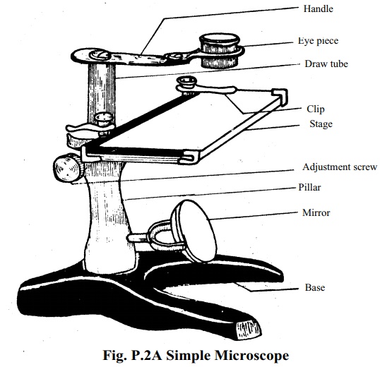
Labelled Diagram of Compound Microscope The below mentioned article provides a labelled diagram of compound microscope. Part # 1. The Stand: The stand is made up of a heavy foot which carries a curved inclinable limb or arm bearing the body tube. The foot is generally horse shoe-shaped structure (Fig. 2) which rests on table top or any other surface on which the microscope in kept.

Compound Microscope: Parts of Compound Microscope
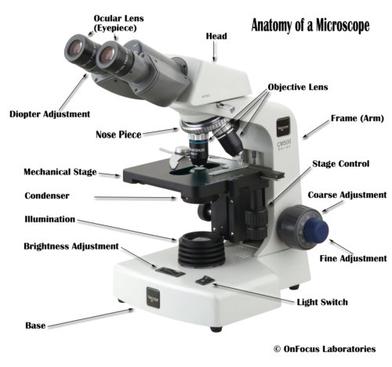
16 Parts of a Compound Microscope: Diagrams and Video Once you have an understanding of the parts of the microscope it will be much easier to navigate around and begin observing your specimen, which is the fun part! The 16 core parts of a compound microscope are: Head (Body) Arm. Base. Eyepiece. Eyepiece tube.

Parts of a Compound Microscope and Their Functions
Simple Squamous Epithelium under a Microscope with a Labeled Diagram ... Simple columnar epithelium labeled. This is a labeled diagram of a simple columnar epithelium under a light microscope. I tried to show you both ciliated and nonciliated simple columnar epithelium. These diagrams show the cilia on the cell surface, rectangular cell, and elongated nucleus.
Simple Microscope - Definition, Diagram, FAQs
assignmentessays.comAssignment Essays - Best Custom Writing Services Get 24⁄7 customer support help when you place a homework help service order with us. We will guide you on how to place your essay help, proofreading and editing your draft – fixing the grammar, spelling, or formatting of your paper easily and cheaply.

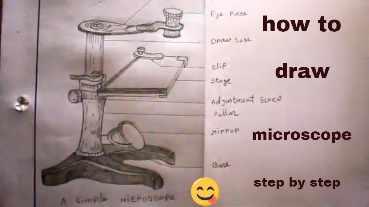
How to draw Microscope diagram for beginners - step by step
Parts of a Microscope Labeling Activity - Storyboard That Create a poster that labels the parts of a microscope and includes descriptions of what each part does. Click "Start Assignment". Use a landscape poster layout (large or small). Search for a diagram of a microscope. Using arrows and textables label each part of the microscope and describe its function. Copy This Storyboard* More options

Free Microscope Drawing, Download Free Microscope Drawing png ...
Microscope Labeling - The Biology Corner Students label the parts of the microscope in this photo of a basic laboratory light microscope. Can be used for practice or as a quiz. ... The type of microscope used in most science classes is the _____ microscope. 18. You should carry the microscope by the _____ and the _____. 19. The objectives are attached to what part of the microscope ...
Microscope: Structure, Uses, Functioning Processes of Simple ...
A Study of the Microscope and its Functions With a Labeled Diagram ... To better understand the structure and function of a microscope, we need to take a look at the labeled microscope diagrams of the compound and electron microscope. These diagrams clearly explain the functioning of the microscopes along with their respective parts. Man's curiosity has led to great inventions. The microscope is one of them.
Microscope Drawing - How To Draw A Microscope Step By Step
Free Microscope Worksheets for Simple Science Fun for Your Students 1. Parts of a Microscope . The first worksheet labels the different parts of a microscope, including the base, slide holder, and condenser. If you have a microscope, compare and contrast this worksheet to it.Also, your kids can color this microscope diagram in and read the words to each part of the microscope.

Dissecting Stereo Microscope Parts and Functions
Simple Microscope Definition, Magnification, Parts And Uses - BYJUS Following are the parts of the simple microscope with their functions: Eyepiece: It is the lens that is used to study the samples and is placed at the top. It has a magnification of 10X to 15X. Base: This provides support to the microscope. Tube: This is used to connect the eyepiece to the objective lenses.

Simple Microscope: Definition, working, diagram, properties, Uses
› createJoin LiveJournal Password requirements: 6 to 30 characters long; ASCII characters only (characters found on a standard US keyboard); must contain at least 4 different symbols;

Parts of a Microscope with Their Functions – Microbe Online
Microscope Drawing Easy with Label - YouTube In this video I go over a microscope drawing that is easy with label. There is a blank copy at the end of the video to review on your own. A great way to s...

Free Microscope Drawing, Download Free Microscope Drawing png ...
PDF Parts of a Microscope Printables - Homeschool Creations Label the parts of the microscope. You can use the word bank below to fill in the blanks or cut and paste the words at the bottom. Microscope Created by Jolanthe @ HomeschoolCreations.net. Parts of a eyepiece arm stageclips nosepiece focusing knobs illuminator stage objective lenses
(159).jpg)
Microscope Quiz: How Much You Know About Microscope Parts And ...
Simple Microscope - Parts, Functions, Diagram and Labelling Parts of the optical parts are as follows: Mirror - A simple microscope has a plano-convex mirror and its primary function is to focus the surrounding light on the object being examined. Lens - The biconvex lens is placed above the stage and its function is to magnify the size of the object being examined.

Parts of a Microscope Labeling Activity
Microscope Labeling - The Biology Corner Microscope Labeling. Shannan Muskopf May 31, 2018. This simple worksheet pairs with a lesson on the light microscope, where beginning biology students learn the parts of the light microscope and the steps needed to focus a slide under high power. The labeling worksheet could be used as a quiz or as part of direct instruction where students label the microscope as you go over what each part is used for.

Simple Microscope- Definition, Principle, Magnification ...
A Study of the Microscope and its Functions With a Labeled Diagram ... May 21, 2019 - To better understand the structure and function of a microscope, we need to take a look at the labeled microscope diagrams of the compound and electron microscope. These diagrams clearly explain the functioning of the microscopes along with their respective parts.

Simple doodles, Microscope parts, Biology labs
Parts of a microscope with functions and labeled diagram - Microbe Notes Head - This is also known as the body. It carries the optical parts in the upper part of the microscope. Base - It acts as microscopes support. It also carries microscopic illuminators. Arms - This is the part connecting the base and to the head and the eyepiece tube to the base of the microscope.

Free art print of Microscope
Parts of the Microscope with Labeling (also Free Printouts) 5. Knobs (fine and coarse) By adjusting the knob, you can adjust the focus of the microscope. The majority of the microscope models today have the knobs mounted on the same part of the device. Image 5: The circled parts of the microscope are the fine and coarse adjustment knobs. Picture Source: bp.blogspot.com.

The Microscope
Microscope Types (with labeled diagrams) and Functions Has a higher level of magnification - Typically up to 2000x. Is used in hospitals and forensic labs by scientists, biologists and researchers to study micro organisms. Compound microscope labeled diagram. Compound microscope functions: It finds great application in areas of pathology, pedology, forensics etc.

4 Best Microscope Drawing and Coloring Pages | Desenhos para ...
Microscope With Labels clip art | Microscope parts, Scientific method ... Description Use this blank handout as a way for students to record microscope drawings. Aside from the drawig itself, studnts are prompted to title the drawing, include the magnification of the microscope, and give a quick description of what they are viewing. Keywords: Microscope Drawings Lab Biology. S. Learning with Shedd Aquarium.
Parts of a Microscope | Microscope Parts and Functions | Labkafe
Label the Microscope Diagram | Download Scientific Diagram - ResearchGate Gram staining was performed using compound microscope according to the procedure described by Petersen et al., 2016 [8]. The gram positive and gram negative bacteria were identified based on ...

Labelling a Microscope Diagram | Quizlet
Parts of a Simple Microscope - Labeled (with diagrams) image 5: A modern simple microscope with the different parts labeled. image source: laboratoryinfo.com The optical parts of a simple microscope are centered on the specimen - lighting, and magnification.
Microscope Parts and Functions

How to Draw a Microscope - Really Easy Drawing Tutorial

How to Draw a Simple Microscope Diagram

Elementary Microscope Parts Poster | Microscope parts ...

Compound Microscope Parts – Labeled Diagram and their ...
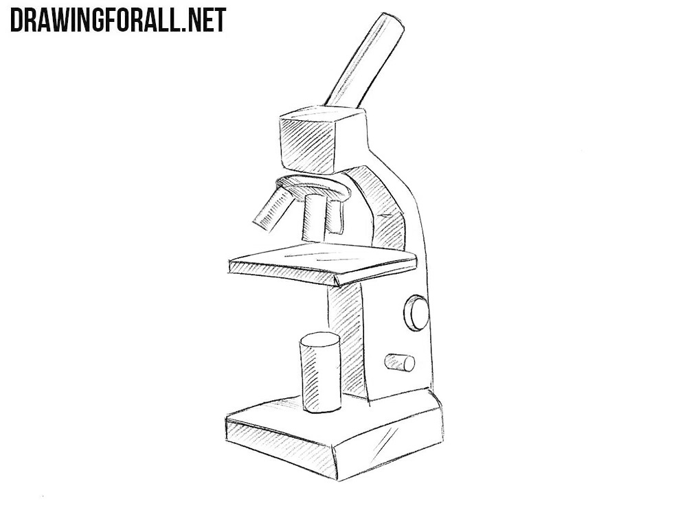
How to Draw a Microscope Easy

Label the microscope — Science Learning Hub
Free Microscope Drawing, Download Free Microscope Drawing png ...

Microscope With Labels Clip Art at Clker.com - vector clip ...
How to Use the Microscope
![How To Draw A Microscope Step by Step - [12 Easy Phase]](https://easydrawings.net/wp-content/uploads/2021/01/Overview-for-Microscope-drawing.jpg)
How To Draw A Microscope Step by Step - [12 Easy Phase]
Free Microscope Drawing, Download Free Microscope Drawing png ...

How to Choose the Perfect Student Microscope — BioBox Labs

Microscope Components - Science Quiz

Label the microscope — Science Learning Hub
How TO Draw simple microscope step by step/simple microscope drawing/for science project

How to draw compound of Microscope easily - step by step
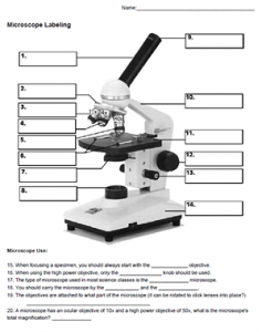
Microscope Labeling

Compound Microscope Parts, Functions, and Labeled Diagram ...

Compound Microscope Parts Made Easy

A Study of the Microscope and its Functions With a Labeled ...
Post a Comment for "42 easy microscope diagram with labels"